The craniomaxillofacial reconstructive surgery was one of the first areas where the 3D printed implants have been used in practice. Modeling has been used in craniofacial reconstruction of traumatic injuries, congenital disorders, orthognathic surgery, tumor removal, and implantology. 3D printing greatly improves and enables preoperative planning, reduce operative time, and significantly improve the biofunctional and the aesthetic outcome.
Computer-aided design (CAD) used to generate 3D proposed implant has a major impact on success of cranio-maxillofacial (CMF) surgery, significantly improving efficiency, accuracy, creativity, and reproducibility.
We care about each patient and provide end-to-end solution. Our highly skilled specialists will analyze 3D image data, design a 3D model of the implant and propose this model to doctors. After satisfying all patient-specific implants’ requirements and getting the approval from doctors we produce the final implant using advanced 3D printing or CNC machining technologies.
The implant and the bone is being printed (plastic). The physical samples is provided to doctor. After feedback and correction the implant is being printed
The doctor has time for pre-operation training and optimization of the surgery process. Then surgery is being performed
Cases
Case 1
Case 1: Orbital flaw: Sphenoid bone
The patient is a Krokodil drug user. Previously he was more than ten times operated on due to osteonecrosis of the maxilla and midface. The result was a severe defect of the midface and the
inferior wall of the orbit. This led to double vision because of the dislocation of one eye. We suggested several models of the implant. However, considering the health condition, quality of bone the doctors chose the version of the implant shown below. The surgery was successful. The main task of recovering the vision as well as the esthetics of the face was accomplished
Process of the work
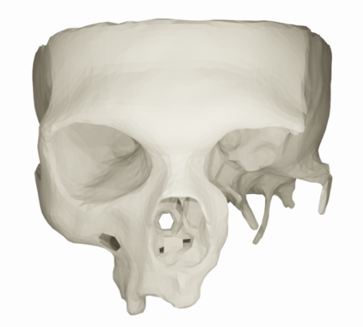
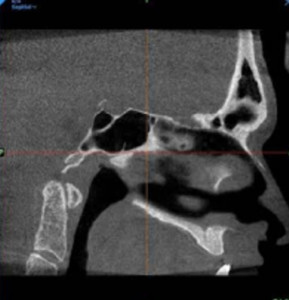
1. Damaged part estimation
Computer tomography
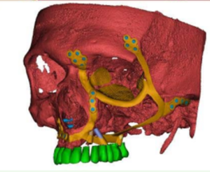
3D model of the damaged bone
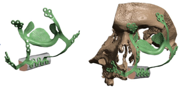
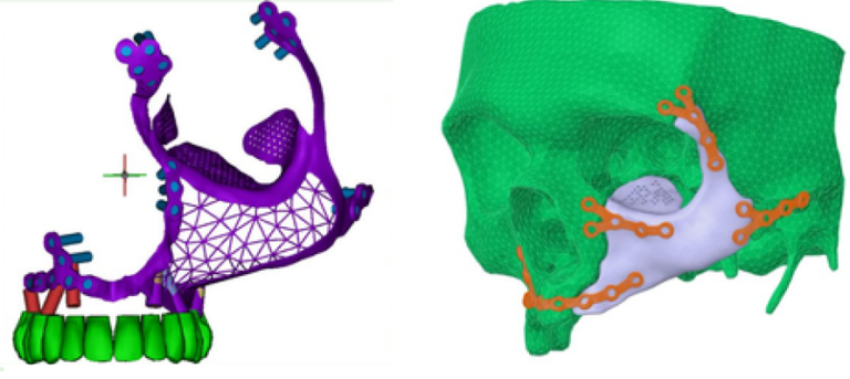
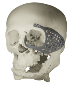
2. 3D models of implants suggested by our medical engineers
Implant model (option 1)
Implant model (option 2)
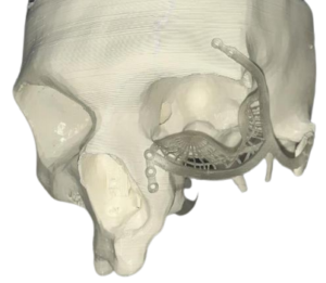
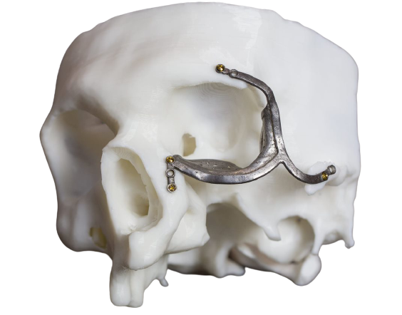
2. Preoperetional training and surgery
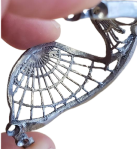
3D printed Titanium implant
Implant assembled with damaged bone
Surgery
Case 2
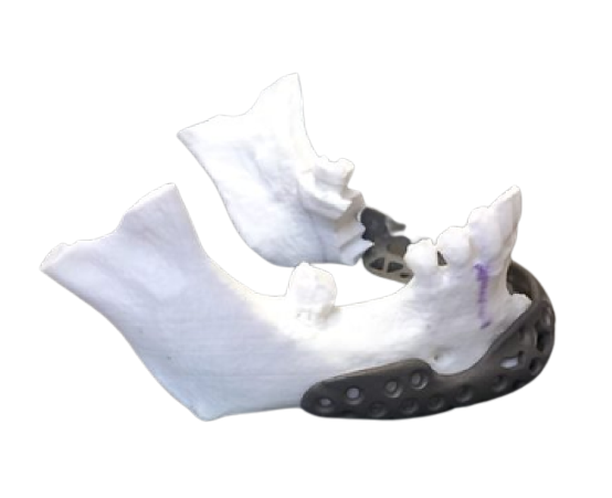
Case 1: Orbital flaw: Mandible implant
The patient presented with Ameloblastoma (arare, non-cancerous benign aggressive large tumor growing towards the jawbone) in the Mandible. Localized from the mental for amenand left body of the Mandible. Cortical resorption and soft tissue infiltration were observed as well. Mandible resection from 43till left angle was planned, and reconstruction with 3D printed mandible implant and vascularized fibula graft was placed.
Process of the work
1. Damaged part estimation
Computer tomography
3D model of bone
2. 3D models of implants suggested by our medical engineers
_ – Mimics Medical 21.0 12_15_2021 2_47_40 PM")